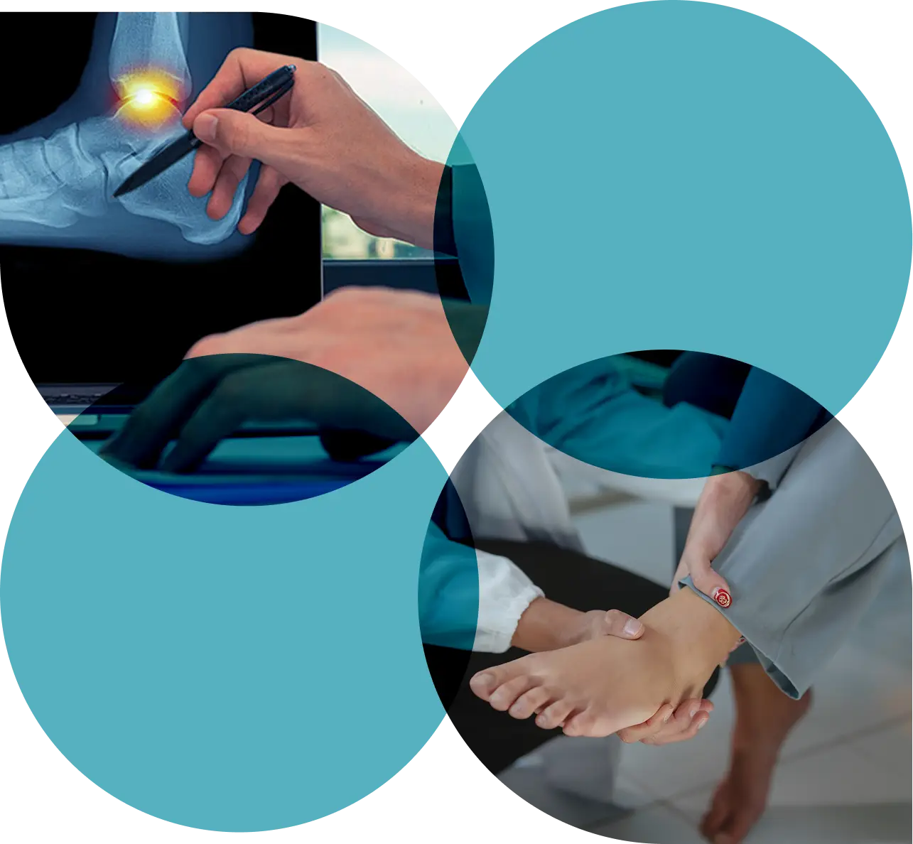
If you have deep ankle pain, swelling, or a catching or locking sensation that hasn't settled since a sprain or injury, you may have an osteochondral lesion. This is where cartilage and bone inside the ankle joint are damaged. Caught early, many lesions respond well to non-surgical care. For larger or unstable lesions, minimally invasive keyhole surgery can relieve symptoms and protect the joint.
Understanding Your Pain
An osteochondral lesion is damage to the cartilage and the bone just beneath it. It's most often found on the talus, which is the bone that forms the base of the ankle joint. It usually follows an ankle sprain or fracture that seemed to heal at the time. The twisting injury bruises or shears the joint surface, and bone and cartilage fragments can loosen rather than heal cleanly.
The condition sits on a spectrum. A small, stable lesion can be symptom-free for years and is often found by chance on a scan. A larger or unstable lesion is more likely to cause persistent deep ankle pain, swelling, or a catching and locking sensation, especially with weight-bearing activity or sport.
If you have already been told you may have an osteochondral lesion, or you are simply tired of ankle pain that has not settled since an old sprain, a private consultation means you can be assessed, diagnosed and started on a treatment plan quickly. For patients with private medical insurance, most treatment, including imaging, injections and surgery, is covered. Self-funding patients are seen at short notice across four central London clinic locations.

Symptom Checklist
Symptoms vary depending on the size and stability of the lesion. Tick anything that applies to you. The more that apply, or the longer they have been present, the more important it is to get a proper assessment.
If three or more of these sound familiar, you are probably overdue for a proper assessment. The earlier we catch an osteochondral lesion, the more treatment options are available to you. Non-surgical approaches work best before the lesion enlarges or becomes unstable.
Treatment Pathway
Most people with an osteochondral lesion do not need surgery. The right treatment depends on the lesion's size, depth and stability, your activity level, and what you have already tried. At every stage, the goal is to use the least invasive approach that reliably relieves your pain and protects the joint. We escalate only when the evidence calls for it.

For small, stable lesions, non-surgical treatment is the starting point and is all many patients need. A plan is built around your specific symptoms and activity level. It typically includes a period of protected weight-bearing or immobilisation, activity modification, and targeted physiotherapy.
Protected weight-bearing
Targeted physiotherapy
Activity modification

When non-surgical care provides partial but insufficient relief, or when the joint is inflamed, an image-guided injection can offer targeted pain relief and help restore function.
Corticosteroid injection
Hyaluronic acid injection
Ultrasound or X-ray guidance

Surgery is considered when non-surgical management has not provided sufficient relief. The choice of procedure depends on the lesion's size and depth, its location, and your activity demands. Mr Malagelada performs every procedure himself.
Arthroscopic bone marrow stimulation
Fragment fixation
Cartilage grafting

LMS, PhD. Consultant Trauma and Orthopaedic Surgeon. Foot and Ankle Specialist.
Consultant at The Royal London
Board member, GRECMIP
Honorary Senior Lecturer
Mr Malagelada is a fellowship-trained Consultant Orthopaedic Surgeon specialising exclusively in conditions of the foot and ankle. He trained in Spain and the UK, including at The Royal National Orthopaedic Hospital and The Royal London, and holds a BOFAS-accredited Foot and Ankle fellowship. He has been a Consultant at The Royal London Hospital since 2017, where he also teaches on the MSc in Orthopaedic Trauma Sciences.

Patient Stories
Real outcomes, independently verified on Doctify.
While the ankle itself might already be uncomfortable, non-surgical treatment such as physiotherapy and activity changes should not be painful. Surgery is carried out under regional or general anaesthetic, so you should not feel the procedure itself. Some soreness and swelling afterwards is normal and is managed with pain relief.
Most arthroscopic procedures take around 45 minutes to just over an hour. The exact time depends on the lesion's size and the technique used, such as bone marrow stimulation, fragment fixation or cartilage grafting.
Arthroscopic, or keyhole, techniques use a small number of tiny incisions, typically a few millimetres each. These usually fade to faint marks. Some scarring is normal with any surgical incision, however small.
Most patients begin putting weight on the affected foot in a controlled way within 2 to 6 weeks. Normal daily activity can usually be resumed by around 3 months. Return to higher-impact sport can take longer and is guided by your physiotherapist and Mr Malagelada.
Yes, many smaller and more stable lesions may never need surgery. Non-surgical care typically includes protected weight-bearing or immobilisation, targeted physiotherapy, and activity modification. Surgery is usually reserved for lesions that stay symptomatic despite a proper trial of this approach.
Mr Malagelada is recognised by Bupa, AXA Health, Aviva, Vitality, WPA and Cigna. If you are self-funding, finance plans over 1 to 5 years are available through Chrysalis Finance.
Still have questions?
Send a question using the form below. A member of the team will reply within one working day.